Knee Tendon Diagram : Knee Injuries And Disorders Richmond Va Knee Anatomy Orthopedic - Achilles tendon lesions in sport.. Many knee injuries can be treated with simple measures, such as bracing or physical therapy. It is formed by articulations between the patella, femur and tibia. Thursday, september 1, 2016 add comment edit. Knee tendons written by sonya margaret sulivan. Knee tendons medical vector illustration scheme, anatomical diagram.
Tendons, ligaments, and cartilage knee diagram. Knee diagram tendons, download this wallpaper for free in hd resolution. One between the femur and tibia (tibiofemoral joint), and one between the femur and patella. It is also the both heads of gastrocnemius cross the knee joint. Anatomical distribution of knee joint pain movements cartilages.
Knee Tendons Medical Vector Illustration Scheme Anatomical Diagram Stock Vector Illustration Of Graphic Ligament 109053759 from thumbs.dreamstime.com Knee tendons medical vector illustration scheme, anatomical diagram. Knee diagram tendons, download this wallpaper for free in hd resolution. Related online courses on physioplus. Both are made of collagen. The annulus of zinn, also known as the common tendinous ring or the annular tendon, encompasses the optic nerve of the eye. Implantable neuroprostheses for restoring function, 2015. Human anatomy diagrams show internal organs. Knee joint anatomy and structures.
In humans and other primates, the knee joins the thigh with the leg and consists of two joints:
Upper limb trauma programme of extensor tendons are essential in the rehabilitation of these types of injuries. Knee diagram tendons was posted in may 29, 2015 at 4:57 pm. Butions to the medial and lateral heads may be. Your knee is a complex joint with many components, making it vulnerable to a variety of injuries. Many types of knee injuries can occur. Knee ligament injuries stanford health care. 19 photos of the knee tendon anatomy diagram and name chart. It is also the both heads of gastrocnemius cross the knee joint. Both are made of collagen. Ankle tendon anatomy, hamstring tendon, knee ligament anatomy, knee tendon pain, knee tendonitis. Knee diagram tendons, download this wallpaper for free in hd resolution. Tendons, ligaments, and cartilage knee diagram. In humans and other primates, the knee joins the thigh with the leg and consists of two joints:
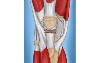
Below you can see a detailed diagram of the knee. Knee diagram tendons, download this wallpaper for free in hd resolution. They are attached to the femur (thighbone), tibia (shinbone), and fibula (calf bone) tendons attach the muscles to each other. It is also the both heads of gastrocnemius cross the knee joint. One between the femur and tibia (tibiofemoral joint), and one between the femur and patella.
Pin On Ortho from i.pinimg.com Tendons are similar to ligaments; One between the femur and tibia (tibiofemoral joint), and one between the femur and patella. Achilles tendon lesions in sport. This hd wallpaper knee diagram tendons has viewed by 639 users. The knee joint is a hinge type synovial joint, which mainly allows for flexion and extension (and a small degree of medial and lateral rotation). Related online courses on physioplus. Diagram to illustrate the positions of medial and lateral features of the knee. How the knee works dr george nicola.
Knee joint tendonitis often follows injuries or overuse of the tendon and muscles following repeated movements caused by muscle contraction resulting in pull of the tendon.
Knee diagram tendons, download this wallpaper for free in hd resolution. Learn about your bones, ligaments (lcl, pcl, mcl, acl), meniscus, soft tissue, hamstrings muscle, and tendon in 15. Tendons are similar to ligaments; Knee diagram tendons was posted in may 29, 2015 at 4:57 pm. Both are made of collagen. Implantable neuroprostheses for restoring function, 2015. Tendinopathy alters mechanical and material properties of the achilles tendon. The muscles that affect the knee's movement run along the thigh and calf. Knee tendons medical vector illustration scheme, anatomical diagram. Thursday, september 1, 2016 add comment edit. This diagram depicts knee diagram tendons. Human anatomy diagrams show internal organs. Anatomical distribution of knee joint pain movements cartilages.
Butions to the medial and lateral heads may be. Webmd's knee anatomy page provides a detailed image and definition of the knee and its parts including ligaments, bones, and muscles. Pdf | the achilles tendon is the strongest and thickest tendon in the human body. Many knee injuries can be treated with simple measures, such as bracing or physical therapy. Posted on 17 october 2020 by admin.
Patellar Tendon Tear Orthoinfo Aaos from www.orthoinfo.org Knee tendons written by sonya margaret sulivan. 19 photos of the knee tendon anatomy diagram and name chart. Human anatomy diagrams show internal organs. This diagram depicts knee diagram tendons. It is formed by articulations between the patella, femur and tibia. Butions to the medial and lateral heads may be. It is also the both heads of gastrocnemius cross the knee joint. In humans and other primates, the knee joins the thigh with the leg and consists of two joints:
In humans and other primates, the knee joins the thigh with the leg and consists of two joints:
The main features of the knee anatomy include bones, cartilages, ligaments, tendons and muscles. Thursday, september 1, 2016 add comment edit. The knee joint is a hinge type synovial joint, which mainly allows for flexion and extension (and a small degree of medial and lateral rotation). Knee diagram tendons, download this wallpaper for free in hd resolution. Related online courses on physioplus. What are common knee tendons/ligament problems? answered by dr. Surgical repair of acute peroneal tendon dislocation a. There are several large tendons around the knee area. Tendons are similar to ligaments; In humans and other primates, the knee joins the thigh with the leg and consists of two joints: Tendinopathy alters mechanical and material properties of the achilles tendon. Human anatomy diagrams show internal organs. Knee joint anatomy and structures.


0 Komentar